A Picture Worth More Than a Thousand Words

Transform the way you see, work and diagnose with the Scopio’s Full Field-PBS™ Application, a standardized approach to enhanced cell visibility.
A Picture Worth More Than a Thousand Words

Digital Cell Morphology Analysis

Discover the potential of real-time, remote collaboration

Maximize Network workflow efficiency
In today’s modern clinical lab, the heavy lifting of the complete blood count with differential (CBC-Diff) is performed by automated hematology analyzers. However, even with advances in hematology analyzer technology, manual examination of peripheral blood smears has been shown to be requested in up to 17% of cases.1 The blood smear provides a visual of the cells present in the bloodstream at any given point in time. Blood cell morphology can add significant value to the routine CBC-Diff, even when the CBC-Diff counts are within the normal ranges.2 Blood cell morphology can be used to look for abnormalities in the shape or quantity of cells;3,4 anemias, cancers and platelet or leukocyte disorders;5,6 or the presence of some blood-borne parasitic infections.7,8

While seemingly simple on the surface, peripheral blood smear evaluation is both time consuming and labor intensive.9,10 Manually evaluating blood smears, even for those with extensive training, can result in significant intra- and inter-operator variability.11 Even with all of the available technology, a skilled morphologist must review all blood smears for morphological review and diagnostic interpretation.12

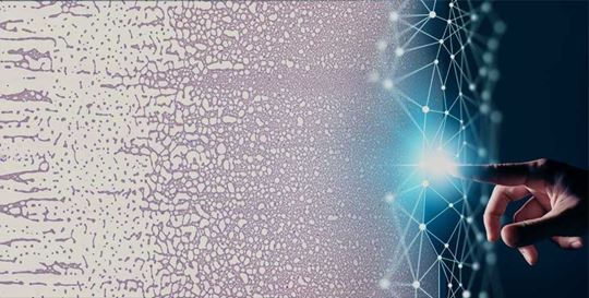
In healthcare, ever-increasing workloads and continually decreasing staffing are putting pressure on laboratory output.13 Clinicians rely on the results of laboratory testing. High laboratorian vacancies due to the pandemic14 and burnout15 have led to current laboratorians being asked to work longer hours, leading to exhaustion and an increased likelihood of errors as well as potential delays in treatment.13,16 Hospital and laboratory networks are primed to consolidate and make more efficient use of their resources, yet fast and easy access to morphology expertise continues to be a challenge—ushering in the need for remote collaboration. Implementing collaborative systems improves efficiency and allows teams to share clinical specialists, both within and outside the hospital’s physical boundaries.

From the monolayer to feathered edge—Viewing what matters
Recent advances in digital hematology have helped to ease the burden on clinical laboratories and cell morphologists. For example, the Slidemaker Stainer Cellular Analysis System automatically prepares slides based on orders received from the integrated hematology analyzer into the work cell and/or from the Laboratory Information System (LIS). 

A well-made blood smear has several parts. The smear base is the thick area where the blood drop was initially placed. Because of its thickness, it dries more slowly than the rest of the slide, which can lead to cellular distortion. This region can be so dense that it becomes difficult to identify individual cells, so it usually isn’t viewed. The middle region of the slide is the monolayer. Because the cells are spread out in the monolayer, it is easier to identify individual cell types. The monolayer is the best place for differential cell counting and cell examination.17 Finally, the feathered edge is found at the end of the smear. Examination of the peripheral blood smear’s feathered edge can aid in the identification of conditions such as pseudothrombocytopenia18,19 and the presence of microfilaria.20

To be Truly Remote, Context Matters
True remote collaboration in hematology digital cell morphology was not available until recently.9,21 In the past, digital cell morphology technology focused on a “snapshot” of a cell, which can provide an accurate, close-up view of a single cell but, because the technology was not able to scan the monolayer to the feathered edge, didn’t provide the cellular context in the patient sample. Personnel specially trained to evaluate blood smears either received a high-resolution image of individual cells with no context or an overview image in low resolution. The morphologist or hematopathologist either had to be physically present in the lab or was only able to look at individual cells. When questions arose with these older systems and context was required, the reviewer had to go back to the manual microscope, which required either going into the laboratory or having the slide sent from a remote location—both of which are inconvenient and time consuming.

With the introduction of Scopio Labs X100 and X100HT scanners, you have a digital image of the clinically relevant part of the smear. Scopio’s Full-Field Digital Cell Morphology™ technology reduces the time9 and tedium associated with manual microscope reviews. And being able to see cells both in full context and zoomed into the smallest detail is vital for confident clinical decision-making.

Clinical-grade AI decision support for insightful peripheral blood smear reviews
Incorporating AI-supported decision making and fully documented digital reporting allows analysis of tens of thousands of cells, leading to faster, more repeatable results—improving every aspect of the hematology workflow and contributing to better patient outcomes.

Scopio’s remote collaboration capabilities increase productivity and efficiency with a simplified and seamless process that facilitates easy communication through digital annotations of the patient sample, replacing chaos with annotatable cases for authorized morphology experts accessing the hospital’s secure network to review—and sign off on—from anywhere.

*Remote review is accessible only for authorized users via the hospital's secure network.

Beckman Coulter is a distributor of Scopio Labs. FDA cleared, CE-marked. Not available in all markets. Please contact your local sales rep for more information about availability.

References:

1. Novis DA, Walsh M, Wilkinson D, St Louis M, Ben-Ezra J. Laboratory productivity and the rate of manual peripheral blood smear review: a College of American Pathologists Q-Probes study of 95,141 complete blood count determinations performed in 263 institutions. Arch Pathol Lab Med. 2006;130(5):596-601. doi:10.5858/2006-130-596-LPATRO

2. Bain BJ. Diagnosis from the blood smear. N Engl J Med. 2005;353(5):498-507. doi:10.1056/NEJMra043442

3. Phillips J, Henderson AC. Hemolytic anemia: evaluation and differential diagnosis. Am Fam Physician. 2018;98(6):354-361.

4. Braunstein E. Megaloblastic Macrocytic Anemias - Hematology and Oncology. Merck Manuals Professional Edition. September 2022. Accessed February 13, 2023. https://www.merckmanuals.com/professional/hematology-and-oncology/anemias-caused-by-deficient-erythropoiesis/megaloblastic-macrocytic-anemias

5. Lynch EC. Peripheral Blood Smear. In: Walker HK, Hall WD, Hurst JW, eds. Clinical Methods: The History, Physical, and Laboratory Examinations. 3rd ed. Butterworths; 1990.

6. Tests for Acute Myeloid Leukemia (AML). American Cancer Society. August 21, 2022. Accessed February 13, 2023. https://www.cancer.org/cancer/acute-myeloid-leukemia/detection-diagnosis-staging/how-diagnosed.html

7. Fernando SD, Ihalamulla RL, Wickremasinghe R, et al. Effects of modifying the World Health Organization standard operating procedures for malaria microscopy to improve surveillance in resource poor settings. Malar J. 2014;13:98. doi:10.1186/1475-2875-13-98

8. Norgan AP, Arguello HE, Sloan LM, Fernholz EC, Pritt BS. A method for reducing the sloughing of thick blood films for malaria diagnosis. Malar J. 2013;12:231. doi:10.1186/1475-2875-12-231

9. Katz B-Z, Benisty D, Sayegh Y, Lamm I, Avivi I. Remote digital microscopy improves hematology laboratory workflow by reducing peripheral blood smear analysis turnaround time. Appl Clin Inform. Published online October 8, 2022. doi:10.1055/a-1957-6219

10. Linder J. www.mlo-online.com/home/article/13004427/digital-imaging-in-hematology. May 1, 2012. Accessed February 7, 2023. https://www.mlo-online.com/home/article/13004427/digital-imaging-in-hematology

11. Palmer L, Briggs C, McFadden S, et al. ICSH recommendations for the standardization of nomenclature and grading of peripheral blood cell morphological features. Int J Lab Hematol. 2015;37(3):287-303. doi:10.1111/ijlh.12327

12. Kratz A, Lee S-H, Zini G, et al. Digital morphology analyzers in hematology: ICSH review and recommendations. Int J Lab Hematol. 2019;41(4):437-447. doi:10.1111/ijlh.13042

13. White R. Laboratory automation is no longer optional. July 24, 2018. Accessed November 17, 2022. https://www.mlo-online.com/information-technology/automation/article/13017029/laboratory-automation-is-no-longer-optional

14. Nuñez-Argote L, Baker DP, Jones AP. Initial Clinical Laboratory Response to COVID-19: A Survey of Medical Laboratory Professionals. Lab Med. 2021;52(4):e115-e124. doi:10.1093/labmed/lmab021

15. Nowrouzi-Kia B, Dong J, Gohar B, Hoad M. Factors associated with burnout among medical laboratory professionals in Ontario, Canada: An exploratory study during the second wave of the COVID-19 pandemic. Int J Health Plann Manage. 2022;37(4):2183-2197. doi:10.1002/hpm.3460

16. Beckering R. The lab shortage crisis: a practical approach | Medical Laboratory Observer. Medical Laboratory Observer. June 1, 2003. Accessed February 13, 2023. https://www.mlo-online.com/home/article/13002441/the-lab-shortage-crisis-a-practical-approach

17. Blood smear features | eClinpath. 2020. Accessed February 9, 2023. https://eclinpath.com/atlas/hematology/blood-smear-features/

18. Nakashima MO. Using Digital Microscopy in Hematology Labs | AACC.org. Clinical Laboratory News. June 1, 2020. Accessed February 8, 2023. https://www.aacc.org/cln/articles/2020/june/using-digital-microscopy-in-hematology-labs

19. CDC - DPDx - Diagnostic Procedures - Blood Specimens. October 1, 2020. Accessed February 9, 2023. https://www.cdc.gov/dpdx/diagnosticprocedures/blood/specimenproc.html

20. Burton E. 6.2 Blood Smear Technique and Reticulocyte Counting. In: CLINICAL VETERINARY DIAGNOSTIC LABORATORY. ; 2021. Accessed February 13, 2023. https://pressbooks.umn.edu/cvdl/chapter/module-7-2-blood-smear-procedure/

21. Katz B-Z, Feldman MD, Tessema M, et al. Evaluation of Scopio Labs X100 Full Field PBS: The first high-resolution full field viewing of peripheral blood specimens combined with artificial intelligence-based morphological analysis. Int J Lab Hematol. 2021;43(6):1408-1416. doi:10.1111/ijlh.13681

Ahmed Bentahar, M.D., Ph.D.
Dr. Bentahar is a clinical analysis specialist (pathologist) with more than 30 years of experience in the field of hematology and laboratory medicine. For the past 27 years, he served as a hematology product specialist, hematology product manager, staff applications scientist and senior manager of global scientific marketing at Beckman Coulter.

Related Articles

Bringing the Future of Hematology 100X Closer

Bringing the Future of Hematology 100X Closer

With their end-to-end digital workflow for PBS, Scopio Labs is transforming hematology cell analysis. Learn how our partnership is enhancing clinical workflows and shaping the future of hematology.
Advancements for the Hematology Laboratory

Advancements for the Hematology Laboratory

New and better solutions for the hematology laboratory were at the center of a virtual roundtable, led by CAP TODAY publisher Bob McGonnagle. Here, Linda Garlaus MT ASCP Senior Manager for Hematology Product Management shares her insights.
What is Monocyte Distribution Width (MDW) and What Role Does It Play in the Early Detection of Sepsis?

What is Monocyte Distribution Width (MDW) and What Role Does It Play in the Early Detection of Sepsis?

A large unmet clinical need for the early detection of sepsis exists. MDW is a novel FDA-cleared biomarker intended to meet this need.